Radioactivity and Air Dose Rate
(2023)
QI heard lichens tend to capture cesium. What is the mechanism like? (2)
AA research team led by JAEA identified the parts of lichens’ body where radioactive cesium are distributed, by combining detection of radioactive materials using a special film and observation of tissues using a digital optical microscope.
In addition, the research team conducted molecular simulations, electron microscopy analysis, and other techniques to suggest the mechanism of cesium retention.
The microscopic radioactive cesium mapping technique and the cesium retention mechanism suggested in this research are likely to help in understanding the mechanism of radioactive cesium retention in the forest ecosystem such as mushrooms and wild grass and the chemical forms of radioactive cesium that affect its movement in the environment.

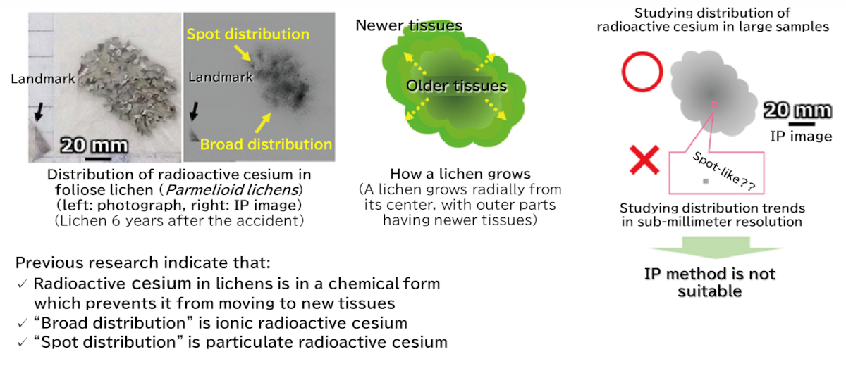
A research team of JAEA, Hosei University, and the National Museum of Nature and Science successfully identified the microscale localization of radioactive cesium in two chemical forms of ions and particles in lichens (lichen = a collective term for a symbiosis between a fungus and an alga) for the first time in the world, finally unveiling the mystery.
The team has been working on the development of a technique for studying the characteristics and spatial distribution of fallout containing radioactive cesium using lichens*1, focusing on the fact that lichens tend to accumulate particulates falling from the atmosphere. Studying such fallout requires demonstrating that radioactive cesium is retained in the body of lichens for a long period of time. However, the mechanism lichens utilize to retain radioactive cesium, that is, “in what part” of their biological tissue radioactive cesium is retained “in what chemical form,” for a long period of time has been a great mystery for many years.
Conventionally, imaging plate autoradiography (IP method)*2 has been used to study two-dimensional distribution of radioactive cesium in environmental samples. However, the IP method has limited resolution (25 μm × 25 μm per pixel), and therefore is not suitable for studying detailed distribution in sub-millimeter ranges. To address the issue, the research team sliced lichens collected in Fukushima prefecture into 5-μm-thick slices using a slicer and took an IP image for each tissue slice to successfully map the distribution of radioactive cesium in much higher resolution of 5 μm than the conventional method. In addition, the team combined the IP images with tissue images taken by a digital microscope*3 to achieve microscale observation of the distribution of radioactive cesium and pigments in tissues.
As a result, it was found as the first case in the world that ionic radioactive cesium*4 was localized in the melanin-like substances (brown pigments)*5 in the lower cortex, while particulate radioactive cesium (radioactive cesium-bearing particles)*6 was localized in the upper cortex to medullary layer parts. Summarizing the localization information as well as results of quantum chemical calculation*7, electron microscopy*8, and other techniques, the team suggested the mechanism of long-term retention of radioactive cesium in lichens as below.
“Melanin substances in the lower cortex may lead to stable complexation*9 of ionic radioactive cesium and contribute to long-term retention. With regard to particulate radioactive cesium, burial on the tissue surface due to development of the mycelia and physical capture into tissues lead to retention of the particles in the upper cortex and in the medullary layer. ”
The microscopic mapping technique established and the retention mechanism suggested in this research are likely to help in the future in elucidating the mechanism of radioactive cesium retention in the forest ecosystem such as mushrooms and wild grass, not just lichens, and the impact of the state of radioactive cesium on its dynamics in the environment.
【Research background and history】
Lichens are commonly seen on land, growing slowly over their long lifetime. They do not have any “root” to absorb substances from; instead, they take in water and other substances through the entire surface of their bodies. For that reason, lichens are known to accumulate substances (radioactive and metal elements including cesium) falling from the atmosphere in their bodies. And for that nature, lichens are recognized throughout the world as a biological indicator (biomonitors) for investigating the level of environmental pollution1). Since the accident at the Fukushima Daiichi NPS (hereinafter simply referred to as the “accident”) that occurred on March 11, 2011, the research team has been working on the development of a technique to study the characteristics and spatial distribution of fallout containing radioactive cesium, focusing on such nature of lichens.
It has been reported to date that (1) the radioactive cesium deposition level immediately after the accident is correlated to the radioactive cesium concentrations in lichens two years after the accident, and radioactive cesium is retained in lichens for a long period of time2), (2) radioactive cesium distribution images taken by the IP method indicate the possibility of radioactive cesium in lichens being retained for a long period of time in a stable chemical form3), and (3) quantum chemical calculation suggests that secondary metabolites produced by lichens form stable complexes with cesium ions depending on the environmental conditions4).
However, “in what parts of the tissue” and “in what chemical forms” lichens in the actual environment retain radioactive cesium for a long period of time have been a mystery for many years.
【Research method】
We focused on some peculiar characteristics of our research subject foliose lichens*1 such as Parmotrema tinctorum, namely, having a thickness of about several hundred μm, having an uncomplicated layered internal structure, and producing different metabolites depending on the layer. The lichen samples used for the research were the “same type” of lichen with “similar level of radioactive cesium,” collected in Fukushima prefecture 2 years and 6 years after the accident, from adjacent locations. We first investigated the trend of radioactive cesium distribution in the whole samples. Then, from the findings of previous research5) 6), we assumed “board distribution” and “spot distribution” to be ionic and particulate radioactive cesium, respectively, and sorted the samples into fragments with both distributions. We then cut the samples into a size of about 1 mm2, horizontally sliced them into a thickness of 5 μm using a slicer, and checked each section for radioactive cesium using the IP method (Figures 1 and 2). Further, for tissue sections identified to have radioactive cesium, we performed extraction processing of IP signal intensity (numeric value) information for each pixel (25 × 25 μm) and colored the pixels by signal intensity range, successfully obtaining a minute-range two-dimensional map of radioactive cesium (Figure 3).
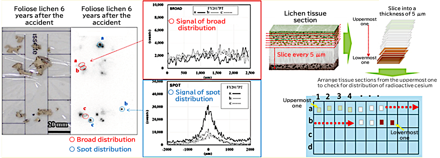
Figure 1 Two-dimensional distribution of radioactive cesium in lichen observed in IP images
A schematic illustration of cutting samples with both “broad distribution” and “spot distribution” into tissue sections.

Figure 2 IP images of tissue sections cut from a fragment with “broad distribution”
Colored frames indicate sections from different tissue layers.
(green : upper cortex and algae, blue : medullary layer, red: lower cortex)
A significant amount of radioactive cesium (white spots in the bottom figure) was detected in some parts of the lower cortex.
【Research results and discussions】
(1) “Broad distribution” in biological tissues of lichens
Commonly in both the lichens collected 2 and 6 years after the accident, it was found that “broad distribution” of radioactive cesium was localized in the lower cortex of the lichens. Further, when an IP image and digital microscope image of sample tissues were combined for each section, as shown in Figure 3, it was shown that the location of brown pigmented tissues was the same as the distribution of radioactive cesium.
Brown pigments in lichens are called “melanin-like substances.” It is possible that radioactive cesium forms stable complexes with these melanin-like substances. To investigate the stability of a complex between a cesium ion and the basic structure of melanin, the representative of melanin-like substances, we employed quantum chemical calculation. As a result, the stability of a complex between a cesium ion and a melanin-like substance was similar to that between a cesium ion and secondary metabolites lichens produce in the upper cortex and medullary layers. However, the IP images of upper cortex and medullary layer tissue sections taken from the actual environmental samples didn’t show any significant amount of radioactive cesium (Figure 2). Meanwhile, it is known that melanin can encapsulate ions of sodium, which is in the same alkali metal group as cesium, using its steric structure5). Therefore, we assumed that the structure of the melanin-like substances in lichens is deeply involved in the long-term retention of ionic radioactive cesium.
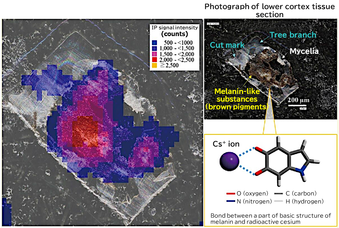
Figure 3 Distribution of radioactive cesium in melanin-like substances in lower cortex tissue section
(2) “Spot distribution” in biological tissues of lichens
Similarly, it was found that “spot distribution” of radioactive cesium was localized around the upper cortex and medullary layer in the tissues of the lichens (Figure 4). Meanwhile, direct electron microscopy observation and elemental analysis indicated the presence of radioactive cesium-bearing particles with sizes of several μm on the surface of the upper cortex to which mycelia adhere densely. Detailed observation of the surface of the upper cortex found that some parts of tissues were adhering to other soil particles of similar size (several μm) and capturing some of them in a buried state (Figure 5). From these findings, we assumed that radioactive cesium-bearing particles adhere to the surface of the upper cortex, then become buried as mycelia grow, resulting in being retained there. Sometimes the surface of the upper cortex develops a crack, which may also cause radioactive cesium-bearing particles to fall into the inside of the medullary layer and become physically captured.
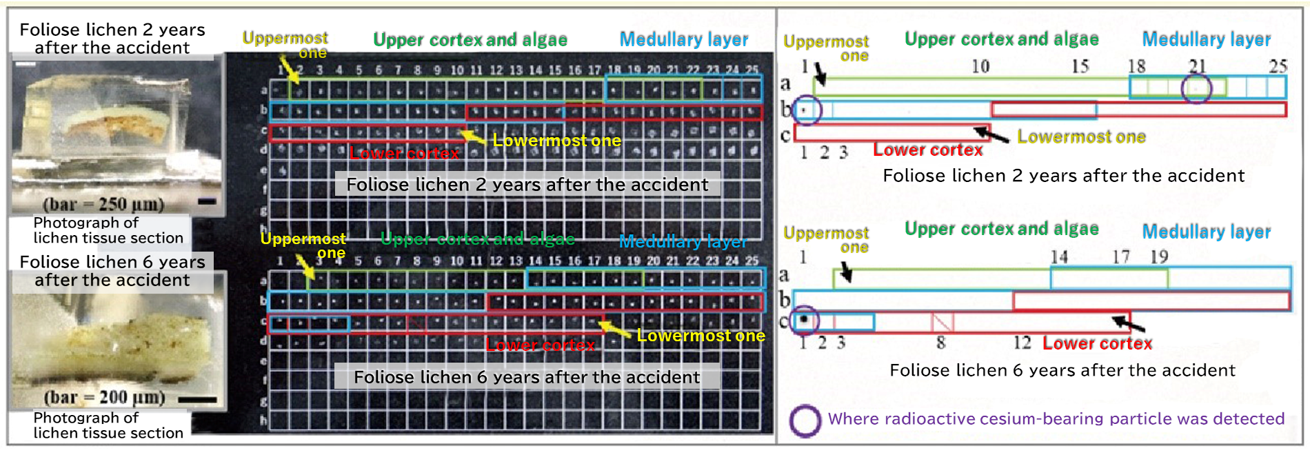
Figure 4 IP images of tissue sections cut from a fragment with “spot distribution”
Colored frames indicate sections from different tissue layers.
(green: upper cortex and algae, blue: medullary layer, red: lower cortex)
The images indicate the presence of radioactive cesium-bearing particles in the medullary or lower cortex layers inside the tissues.

Figure 5 An electron microscopy image of the surface tissue of the upper cortex
Many particles (white spots in the image; primarily soil particles) were found captured.
From these findings, we suggest the mechanism of long-term retention of radioactive cesium that enters into lichens through the atmosphere or rainwater. “Melanin substances in the lower cortex may lead to stable complexation of ionic radioactive cesium and contribute to long-term retention. With regard to particulate radioactive cesium, burial on the tissue surface due to development of the mycelia and physical capture into tissues lead to retention of the particles in the upper cortex and in the medullary layer. ”
【Future perspective】
In this research, we established a series of techniques to investigate detailed microscale distribution and chemical forms of radioactive cesium in biological tissues of lichens which are commonly available in the environment and easy to handle. We believe these techniques are applicable to other environmental samples and will contribute to elucidating “the state of radioactive materials in the natural environment,” instead of relying on hypotheses built through experiments in laboratories. We also discovered the mechanism of retention of radioactive cesium, like melanin-like substances are deeply involved in the long-term retention of ionic radioactive cesium. The microscopic mapping technique established and the retention mechanism suggested in this research are likely to help in the future in elucidating the mechanism of radioactive cesium retention in the forest ecosystem such as mushrooms and wild grass, not just lichens, and the impact of the state of radioactive cesium on its dynamics in the environment.
【Terms】
- *1 Lichens :
Lichen is a collective term for symbiosis between a fungus and an alga. Lichens are organisms commonly found in the terrestrial ecosystems, including human’s living space (e.g., rock surfaces, tree trunk surfaces, house walls, concrete, stonework), mountains, forests, polar regions, and arid areas. They remain in one place while they grow. Their growth speed is slow (approx. several mm per year), and they live relatively long with a lifespan of several dozen years.
Lichens are broadly classified into three types, foliose lichens, crustose lichens, and fruticose lichens, by their appearance. Foliose lichens have a flat, leaf-like thin shape, and they loosely attach to the substrate on which they grow by rhizoids (root-like structures). Crustose lichens appear like tree bark or stone surface patterns. They do not have a lower cortex layer, and their mycelia directly fasten to the substrate on which they grow. Fluticose lichens have a bushy structure, and they grow standing up on or hanging down from the substrate surface.
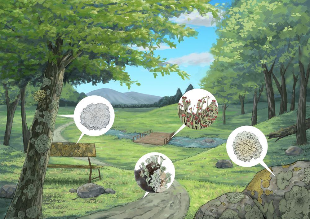
Figure Illustration of lichens growing in the environment
Lichens can be seen in various places around us, such as on trees, the ground, rocks, and bridges. - *2 Imaging plate autoradiography (IP method) :
When an observation sample is placed on a special film sensitive to radiation (= imaging plate, IP) and then the IP is processed by a device to form an image for radiation-sensitized areas, a two-dimensional image showing the position or distribution of the sources of radiation that sensitized the IP can be obtained. Comparing the image with a photograph of the sample enables easy visual understanding of “where radioactive materials are located (distributed) in the sample,” and this “IP method” is widely used. - *3 Digital microscope :
Digitally-processed optical microscope. In the case of a general optical microscope, a sample is observed through an eyepiece. Meanwhile, in the case of a digital microscope, a sample is observed using a camera and images taken by the camera are enlarged and displayed on a monitor. - *4 Ionic radioactive cesium :
Cesium is one of alkali metals (other well-known alkali metals are sodium and potassium), and becomes monovalent cations (positively charged) when dissolved in water. Ionic radioactive cesium refers to this monovalent cation radioactive cesium. - *5 Melanin-like substances (brown pigments) :
Melanin is a (usually black) pigment that is produced in a wide variety of organisms including humans, animals, and plants. Some lichens also have pigments with a basic structure similar to melanin in their tissues, and these pigments are called melanin-like substances. - *6 Particulate radioactive cesium (radioactive cesium-bearing particles) :
Matters that are generally recognized as particles but contain radioactive cesium are called “particulate radioactive cesium” or “radioactive cesium-bearing particles.” Examples include soil mineral particles, metal particles, glass particles, and organic matter aggregates. - *7 Quantum chemical calculation :
A technique to analyze the structure and characteristics of atoms and molecules from their electronic states. It enables calculating at what distance atoms and molecules form a bond, and how strong the bond (energy required for forming the bond) is. Since it is to seek the optimal structure from a huge number of atoms and molecular structure candidates, calculation is performed using large-scale computers. - *8 Electron microscopy :
In optical microscopy, a sample is observed under normal light (visible light). Meanwhile, in electron microscopy, electron beams (which have much shorter wavelengths than visible lights) are irradiated at a sample to observe the sample in a resolution much higher than optical microscopy. By the electron microscope used in this research, elements contained in a sample can be investigated by checking the energy and wavelength of X-rays emitted from the sample irradiated with electron beams. - *9 Complexation :
It refers to formation of a complex (a collective term for molecules created by bonding between an atom and a molecule, between an ion and a molecule, or between different molecules).